| Title | Pages |
|---|---|
| Preface |
Preface |
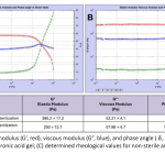
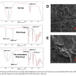
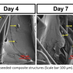
| Regenerative Material for the Treatment of Articular Cartilage Damage: Synthesis and Characterization of Hyaluronic Acid and Poly(glycolic acid) Composite Matrices Currently, analgesics, anti-inflammatory drugs, and hyaluronic acid viscosupplementation are used to alleviate pain associated with joint cartilage disorders. Hyaluronic acid injections are known not only for their pain-reducing effects but also for stimulating cartilage regeneration. In this study, a regenerative biomaterial platform comprising poly (glycolic acid)mesh and cross-linked hyaluronic acid was developed for the repair of degenerated joint cartilage following microfracture and subchondral bone stimulation. For this purpose, in the first stage, hyaluronic acid gels cross-linked with butanediol diglycidyl ether, containing a concentration of 23 mg/mL, were prepared. The residual butanediol diglycidyl ether cross-linker in the obtained gels was below 1 ppb. The pH value was determined to be 6.95 ± 0.2, and the osmolality was 361.3 ± 2.9 mOsm/kg. The injection force and related rheological properties were investigated. In the second stage, the cross-linked hyaluronic acid gels were impregnated into poly (glycolic acid) meshes, evaluated using scanning electron microscopy and characteri-zed chemically. Finally, the composite matrices were recellularized with chondrocytes, and cell viability analysis was conducted using Alamar Blue. The cell viability results and scanning electron microscopy images of the composite structure consisting of poly(glycolic acid) mesh and cross-linked hyaluronic acid indicated that the structure supports chondrocyte viability. 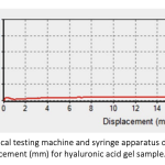
|
379 - 388 |
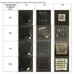
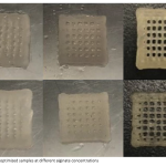
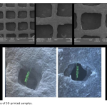
| Development of 3D Printed Scaffolds Containing Decellularized Plants and Investigation of Their Basic Cell Interactions The decellularization process fundamentally removes the cellular content of the tissue (nuclear material and other nucleic acid components) without disrupting the structural integrity of the tissue. It is an effective approach, especially for obtaining three-dimensional (3D) biomaterials composed of the extracellular matrix (ECM), which provides tissue biomechanical support. In the literature, studies have shown that after the decellularization process, animal-derived decellularized tissues have been combined with various biopolymers to prepare composite scaffolds using different techniques. In recent years, due to their structural features, decellularization studies of plant-derived tissues have also gained prominence alongside animal tissues. In this study, succulent plants were chosen as the plant tissue, and the purpose was to prepare hybrid scaffolds by combining decellularized succulent tissues with alginate structures and to investigate the fundamental cell-material interactions using mesenchymal stem cells. Succulent plant leaves were decellularized using a solution containing Triton X-100 and SDS. The water-retaining parts were separated from other tissues, lyophilized, and turned into a powder. This approach was employed to preserve biomolecules with water-retaining capacity in powdered form. To determine the efficiency of the decellularization process, the quantities of DNA and proteins were assessed and compared. Due to their high water-absorbing capacity, the succulent plants' water-retaining structures were combined with alginate biopolymer at various viscosity levels to prepare an ink suitable for 3D printing. After printing, the resulting scaffolds' degradation and swelling behavior, chemical composition, structural characterization, and thermal properties were examined. In the final phase, a fundamental investigation was carried out on cell-material interactions using L929 mouse fibroblast cells and human mesenchymal stem cells on 3D-printed scaffolds. The interactions within the prepared hybrid scaffolds were analyzed through basic cytotoxicity tests. 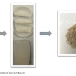
|
389 - 404 |
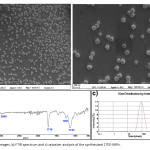
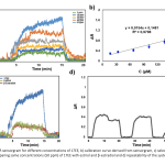
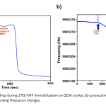
| Harnessing Molecularly Imprinted Polymers in Surface Plasmon Resonance and Quartz Crystal Microbalance for 17α-Ethinyl Estradiol Detection Estradiol is a critical hormone for reproductive health in both females and males, playing a key role in diagnosing conditions such as menopause, infertility, and certain cancers. However, estradiol, especially in its synthetic form, 17α-ethinyl estradiol (17EE), also represents a significant environmental threat as an endocrine-disrupting chemical (EDC), with serious implications for ecosystems and human health. Despite growing awareness of 17EE's role in water contamination and its potential to disrupt aquatic ecosystems and human endocrine systems, existing detection methods for 17EE are limited. Establishing reliable, rapid, and sensitive biosensors for estradiol detection is thus essential for both environmental monitoring and healthcare. In this study, 17EE-imprinted polymeric nanoparticles (17EE-MIPs) were synthesized using mini-emulsion polymerization and characterized. The synthesized 17EE-MIPs were evaluated using Surface Plasmon Resonance (SPR) and Quartz Crystal Microbalance (QCM) platforms. SPR analysis yielded equilibrium and binding kinetic parameters, with the Freundlich model providing the best fit for the 17EE-MIP based system. Following this, 17EE-MIPs were covalently attached to a QCM crystal, where consecutive 17EE concentrations were tested. Both platforms demonstrated high linearity in detecting low concentrations of 17EE, with detection limits of 11.57 µM and 1.335 µM for SPR and QCM, respectively. These results underscore the potential of using MIPs to develop highly sensitive biosensors for hormone detection, with applications in both environmental and healthcare fields. 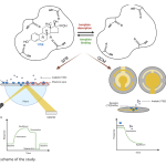
|
405 - 414 |
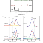
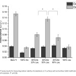
| Amino Acid Conjugated Self Assembled Molecules Modified Titanium Surfaces For Investigating Osteoblast Behavior In this study, human fetal osteoblasts behavior was investigated on titanium surfaces that has been modified with amino acid conjugated self-assembled molecules. For this purpose, 3-aminopropyltriethoxysilane (APTES) was conjugated by histidine and leucine and these newly synthesized molecules were used in different combinations to modify titanium surfaces via creating amino acid conjugated self-assembled monolayers (SAM) on titanium surfaces. The modification of the surfaces to introduce hydrophilic and hydrophobic regions on the surface was achieved with varying concentrations (v/v,100:0 20:80, 50:50, 80:20, 0:100). X-ray photoelectron spectroscopy (XPS) analysis and water contact angle measurements were performed for characterizing all of the modified surfaces in order to verify presence of amino acid specific bonds and wettability behavior to find suitable concentrations to support initial cell adhesion. In order to confirm that the surface modification supported cell adhesion and proliferation, 3-(4,5-dimethylthiazol-2-yl)-2,5-diphenyltetrazolium bromide (MTT) assay was performed. Our results have shown that, amino acid SAM modification can be used to fine tune surface wettability and adherent cells were able to proliferate at different rates using different mixture concentrations. This presented approach can prove useful for expanding fine tuning surface chemistry methods for more specific applications and research. 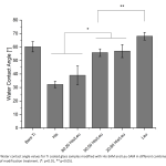
|
415 - 423 |
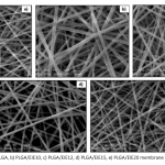
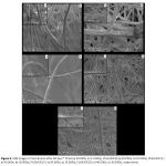
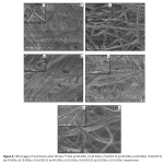
| Production of Polymeric Nanofiber Membranes Containing Plant Extract and Investigation of Their Potential for Use on Wound Healing In cases of skin tissue injury, such as severe burns, skin tissue layers can be extensively damaged. Various dressings have been developed to address different types of wounds, with functional polymeric dressings being the most popular. These advanced dressings are designed to accelerate wound healing. Incorporating plant-derived extracts and biological molecules into wound dressing materials is common. This study aimed to develop an electrospun nanofiber wound dressing by incorporating active ingredients extracted from the plant Echium italicum (Italian viper's bugloss), known for its efficacy in burn wound healing, into poly (lactic-co-glycolic acid) (PLGA). The nanofiber membrane wound dressings produced by electrospinning were subjected to various analyses. Morphological and structural characterization revealed that under physiological conditions, the membranes exhibit significant morphological decomposition and weight loss after 90 days of vitro degradation. In vitro cytotoxicity testing using the MEM extraction method demonstrated that the membranes were not cytotoxic. Based on the comprehensive analysis, it was concluded that the developed nanofiber membranes hold promise as potential wound dressings for the treatment of severe burn wounds. 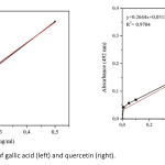
|
425 - 437 |